 Most people come in knowing something hurts. They do not always know why.
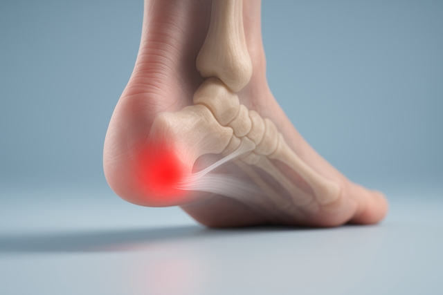
A knee that aches when going downstairs might be cartilage wear. It might be that the patella is tracking incorrectly. It might be referred pain from the hip. A shoulder that hurts overhead might be impingement, a rotator cuff tear, or a cervical spine issue producing arm pain that has nothing to do with the shoulder joint itself.
Pain tells you something is wrong. It does not tell you what. That is what the evaluation is for.
Medical History
The first part of any evaluation is understanding the full picture before anything gets examined physically. How long has the pain been present? What happened around the time it started? Whether it came on suddenly or built up over weeks. What makes it better and worse? Whether it travels anywhere.
Previous injuries and surgeries matter. What has already been tried and whether it helped. These details shape the clinical picture before anyone touches the patient.
Physical Assessment
Range of motion gets tested in the affected joint and the ones above and below it. Strength testing identifies which muscles are not supporting or controlling the area properly. Palpation locates where tenderness sits and what reproduces the pain. Neurological testing comes in when nerve involvement is possible. Specific stress tests load particular structures to identify which ones are contributing.
Two patients with identical pain locations can have completely different findings. One has a mobility restriction, driving the problem. The other has a strength deficit. The treatment looks different for each.
Movement Analysis
Static testing only tells part of the story. Watching how the body moves under real conditions reveals the rest.
A squat that shows the knee caving inward under load. A shoulder elevation relying on the upper trap because the lower scapular stabilizers have checked out. A walking pattern that consistently offloads one side. These movement faults put uneven stress on specific structures across hundreds of repetitions and are often why pain keeps returning despite treatment aimed directly at the painful site.
Imaging Review
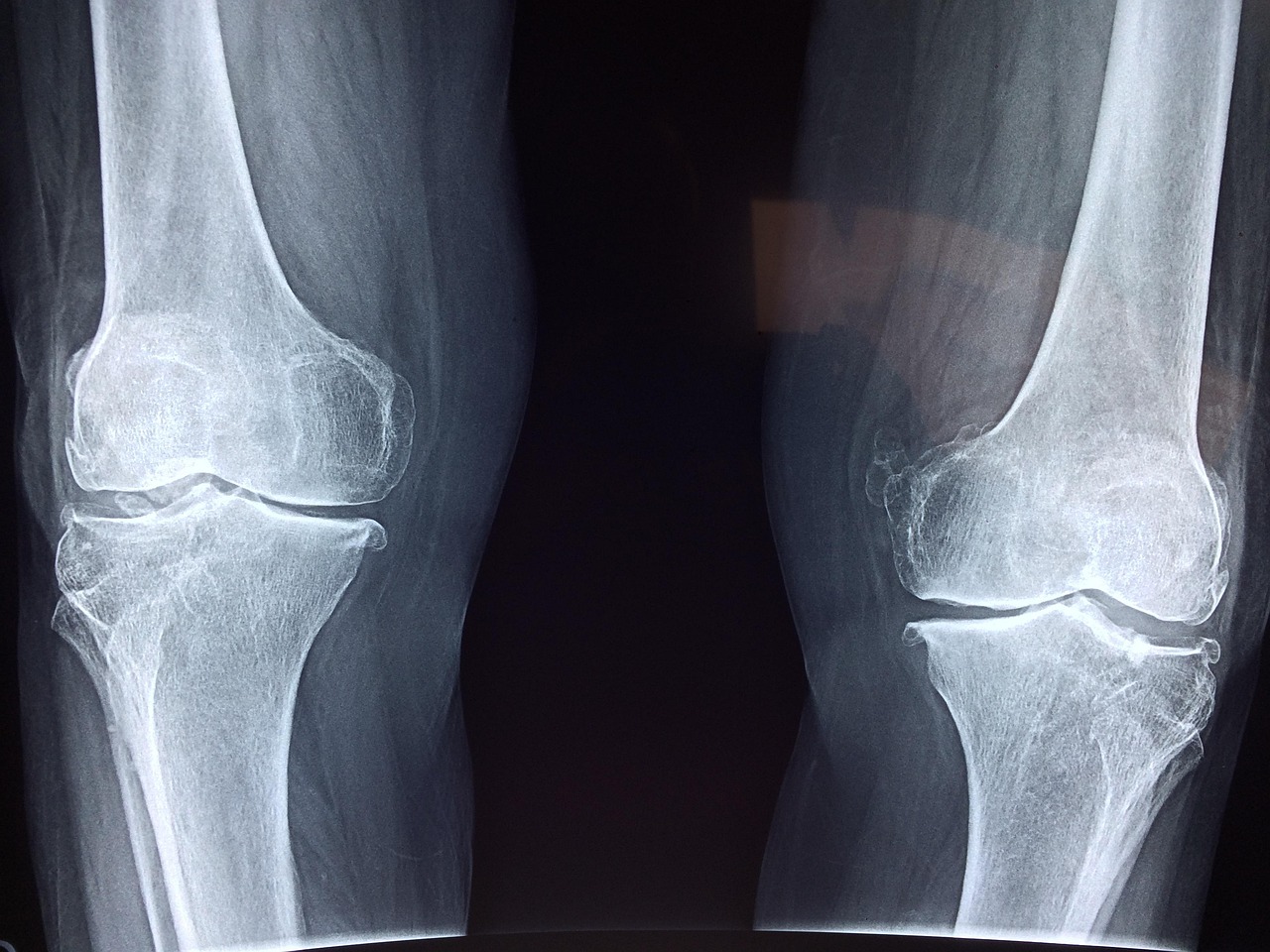
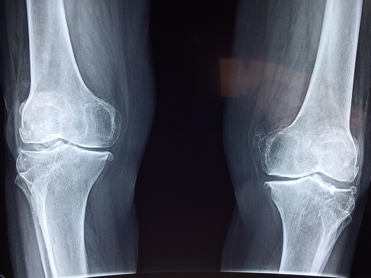
X-rays and MRI show structure but not function. A disc bulge on an MRI may be the source of the pain, or it may be incidental. Cartilage loss on an X-ray tells part of the story, but not how the person is actually moving or which muscles have stopped working around that joint.
Imaging gets interpreted alongside the clinical findings rather than treated as the definitive answer on its own. When imaging has not been done and the clinical picture suggests it would change the plan, the provider points the patient in the right direction.
Let us help. If persistent pain has been limiting daily life, reach out to Modern Medicine of Southeast Tucson, AZ, to get a proper evaluation scheduled.
Most people come in knowing something hurts. They do not always know why.
A knee that aches when going downstairs might be cartilage wear. It might be that the patella is tracking incorrectly. It might be referred pain from the hip. A shoulder that hurts overhead might be impingement, a rotator cuff tear, or a cervical spine issue producing arm pain that has nothing to do with the shoulder joint itself.
Pain tells you something is wrong. It does not tell you what. That is what the evaluation is for.
Medical History
The first part of any evaluation is understanding the full picture before anything gets examined physically. How long has the pain been present? What happened around the time it started? Whether it came on suddenly or built up over weeks. What makes it better and worse? Whether it travels anywhere.
Previous injuries and surgeries matter. What has already been tried and whether it helped. These details shape the clinical picture before anyone touches the patient.
Physical Assessment
Range of motion gets tested in the affected joint and the ones above and below it. Strength testing identifies which muscles are not supporting or controlling the area properly. Palpation locates where tenderness sits and what reproduces the pain. Neurological testing comes in when nerve involvement is possible. Specific stress tests load particular structures to identify which ones are contributing.
Two patients with identical pain locations can have completely different findings. One has a mobility restriction, driving the problem. The other has a strength deficit. The treatment looks different for each.
Movement Analysis
Static testing only tells part of the story. Watching how the body moves under real conditions reveals the rest.
A squat that shows the knee caving inward under load. A shoulder elevation relying on the upper trap because the lower scapular stabilizers have checked out. A walking pattern that consistently offloads one side. These movement faults put uneven stress on specific structures across hundreds of repetitions and are often why pain keeps returning despite treatment aimed directly at the painful site.
Imaging Review
X-rays and MRI show structure but not function. A disc bulge on an MRI may be the source of the pain, or it may be incidental. Cartilage loss on an X-ray tells part of the story, but not how the person is actually moving or which muscles have stopped working around that joint.
Imaging gets interpreted alongside the clinical findings rather than treated as the definitive answer on its own. When imaging has not been done and the clinical picture suggests it would change the plan, the provider points the patient in the right direction.
Let us help. If persistent pain has been limiting daily life, reach out to Modern Medicine of Southeast Tucson, AZ, to get a proper evaluation scheduled.
Green Valley, AZ – What Non Surgical Orthopedic Clinics Review in Pain Exams
SYNOPSIS: This article covers non-surgical orthopedic pain evaluations, including what gets assessed, why each part matters, and how to understand what is actually driving persistent pain.
What an Orthopedic Pain Evaluation Covers
BY: Josh Adams, Modern Medicine of South East Tucson, AZ
Most people come in knowing something hurts. They do not always know why.
A knee that aches when going downstairs might be cartilage wear. It might be that the patella is tracking incorrectly. It might be referred pain from the hip. A shoulder that hurts overhead might be impingement, a rotator cuff tear, or a cervical spine issue producing arm pain that has nothing to do with the shoulder joint itself.
Pain tells you something is wrong. It does not tell you what. That is what the evaluation is for.
Medical History
The first part of any evaluation is understanding the full picture before anything gets examined physically. How long has the pain been present? What happened around the time it started? Whether it came on suddenly or built up over weeks. What makes it better and worse? Whether it travels anywhere.
Previous injuries and surgeries matter. What has already been tried and whether it helped. These details shape the clinical picture before anyone touches the patient.
Physical Assessment
Range of motion gets tested in the affected joint and the ones above and below it. Strength testing identifies which muscles are not supporting or controlling the area properly. Palpation locates where tenderness sits and what reproduces the pain. Neurological testing comes in when nerve involvement is possible. Specific stress tests load particular structures to identify which ones are contributing.
Two patients with identical pain locations can have completely different findings. One has a mobility restriction, driving the problem. The other has a strength deficit. The treatment looks different for each.
Movement Analysis
Static testing only tells part of the story. Watching how the body moves under real conditions reveals the rest.
A squat that shows the knee caving inward under load. A shoulder elevation relying on the upper trap because the lower scapular stabilizers have checked out. A walking pattern that consistently offloads one side. These movement faults put uneven stress on specific structures across hundreds of repetitions and are often why pain keeps returning despite treatment aimed directly at the painful site.
Imaging Review
X-rays and MRI show structure but not function. A disc bulge on an MRI may be the source of the pain, or it may be incidental. Cartilage loss on an X-ray tells part of the story, but not how the person is actually moving or which muscles have stopped working around that joint.
Imaging gets interpreted alongside the clinical findings rather than treated as the definitive answer on its own. When imaging has not been done and the clinical picture suggests it would change the plan, the provider points the patient in the right direction.
Let us help. If persistent pain has been limiting daily life, reach out to Modern Medicine of Southeast Tucson, AZ, to get a proper evaluation scheduled.